
Kolling researcher and physiotherapist Brian Kim has spent most of his career in orthopaedic clinics, helping patients recover from or prepare for surgery.
Over time, he noticed something unusual in MRI scans, some muscles showed streaks or “marbling” of fat. While tears and inflammation were easy to spot, this fatty appearance was harder to interpret.
“I’d look at my patients’ scans and think, I don’t think that’s a good thing,” Brian said. “It’s a very clear feature on imaging, but we just didn’t know how to measure it accurately and therefore, whether it is truly important or not.”
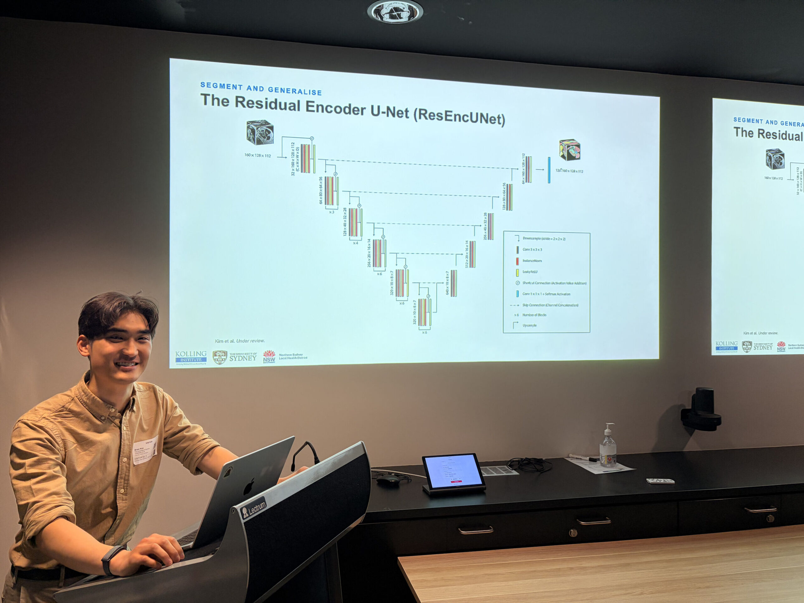
That question sparked a research journey for Brian, which has involved using artificial intelligence (AI) to automate the identification of fat and muscle in MRI scans, focusing on the shoulder.
So far, the PhD candidate at the Kolling Institute has analysed 700 scans using advanced imaging techniques and compared the results to assessments done by hand.
Brians explains that a standard MRI shoulder scan, which shows only part of the shoulder muscles, would usually take about an hour to complete by hand. Full high-resolution scans, often needed before surgery, can take up to six hours.
With AI, the same task takes just seconds for standard MRIs and 50 seconds for detailed CT scans that extend past the elbow.
Why does it matter?
Even a small amount of fat, around seven or eight percent, can increase the risk of poor surgical outcomes. “Although the majority of surgeries are successful, there are opportunities to improve.”
“A surgeon could repair a tendon, and then it tears again within six months to a year,” he said. “In the shoulder, you want very sturdy, muscle-only muscles.”
There are different types of fat in the body – under the skin, around organs, inside and outside muscles. As Brian puts it, “Not all fats are created equal.”
Muscle fat often develops from disuse, such as after injury or long-term pain. For example, people with chronic low back pain or whiplash often develop fatty streaks in their back or neck muscles. And once this fat appears, it’s irreversible – at least for now.
Brian’s goal is to understand the cellular reasons behind muscle fat. If researchers can pinpoint why it forms, they may one day find ways to prevent or reverse it.
“I see muscle tissue as a cornerstone of healthy living. It shows when we’ve had injuries, are fearful of movement and even if we are staying active and high-spirited,” he said.
He’s hopeful that in future, MRI reports will include both radiologist findings and muscle fat data. This could help surgeons make more informed decisions. For instance, if one part of a muscle shows high fat, they may choose to operate only on the healthier section to avoid re-tear.
“It helps us diagnose better, not just the disease, but the severity of muscle degeneration,” Brian said. “And we can monitor recovery more accurately after surgery.”
Ultimately, Brian wants to build a large dataset of muscle volume and fat percentages – a bit like the information gleaned from a blood test – to guide better care.
Brian’s work is helping turn muscle fat from a mystery into a measurable tool for better care.

